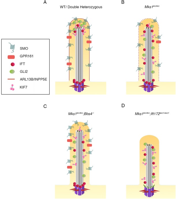
Fig 8. Model summarizing the trafficking defects in Mks1krc/krc mutants as well as double mutants with Bbs and Ift.
All conditions are depicted in the state of HH pathway activation upon stimulation with SAG. (A) WT and double heterozygous cells. Red outline depicts the normal ciliary membrane integrity (based on ARL13b and INPP5E localization) SMO is present in the ciliary membrane, GPR161 is reduced within the ciliary membrane due to pathway activation. GLI2 and KIF7 are enriched at the ciliary tip. IFT components are enriched at the ciliary tip and the transition zone near the basal body (shown in purple). (B) Mks1krc/krc cells exhibit defects in the localization of ciliary membranes (dashed red line)- ARL13b is reduced and INPP5E is absent. SMO is reduced, and GLI2, KIF7, and IFT81 are no longer restricted to the ciliary tip. (C) Mks1krc/krc cells that also lack BBS4 exhibit additional defects in the trafficking of ciliary membrane proteins. ARL13B is absent, however SMO is restored in the cilia by the loss of BBS4. GLI2 is not restricted to the ciliary tip, as with Mks1krc/krc single mutant cells, however a lower percentage of cells have ciliary GLI2. (D) Mks1krc/krc cells that also have impaired IFT function due to a homozygous hypomorphic mutation in Ift172 have severe ciliogenesis defects. Less than 10% of Mks1krc/krc;Ift172avc1/avc1 cells are ciliated. Those cilia that are present are shorter than normal. IFT88 is absent from the transition zone or the cilium, while IFT81 and KIF7 are no longer restricted to the tip, as with Mks1krc/krc single mutant cells.

