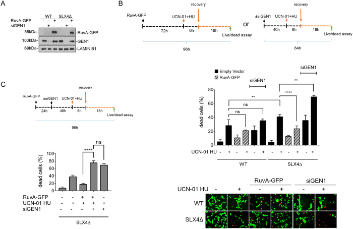
Figure 7. RuvA expression increases viability of FA-P cells after pathological replication stress.
(A) Analysis of RuvA-GFP expression or GEN1 depletion by Western blotting in FA-P and FA-P complemented cells. Immunoblotting was performed using the indicated antibodies. GAPDH was used as loading control. (B) After transfection with RuvA-GFP transfection or siRNAs oligo, as depicted in the experimental scheme, cells were treated with UCN-01 (600 nM) and HU (2 mM) and analyzed by LIVE/DEAD assay. Data are presented as percentage of dead cells and are mean values from three independent experiments. Error bars represent standard errors (SE). (ns = not significant p > 0.05; **p < 0.01, ****p < 0.0001, Mann-Whitney test). The panel shows representative images: live cells are green stained, while dead cells are red. (C) GEN1 depletion counteracts RuvA expression. Twenty-four hours after transfection with RuvA-GFP, FA-P cells were transfected with GEN1 siRNAs (siGEN1) and 24 h thereafter cells were treated with UCN-01 (600 nM) and HU (2 mM) for 6 h, according to the experimental scheme. After the recovery, cells were analyzed by LIVE/ DEAD assay. Data are presented as percentage of dead cells and are mean values from three independent experiments. Error bars represent standard errors. (ns = not significant p > 0.05; **p < 0.01, ****p < 0.0001, Mann-Whitney test). Uncropped versions of gels are provided in Supplementary Material.

