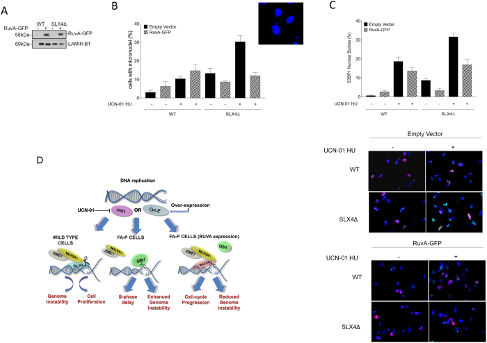
Figure 8. GEN1 premature activation causes genome instability in absence of SLX4.
(A) Western blotting analysis showing expression of RuvA-GFP in FA-P-derived cells. (B) Analysis of micronuclei (MN). Forty-eight hours after RuvA-GFP transfection, FA-P and FA-P complemented cells were treated with 600 nM UCN-01 and 2 mM HU for 6 h, recovered overnight in drug-free medium and analysed for the presence of MN by DAPI staining. Data are presented as mean values of percentage of cells with MN from three independent experiments. Error bars represent standard errors. The panel shows representative images of micronuclei-containing FA-P cells after replication stress. (ns = not significant p > 0.05; **p < 0.01, ****p < 0.0001, Mann-Whitney test). (C) 53BP1 NBs formation in FA-P and FA-P complemented cells. After RuvA-GFP transfection, cells were treated with 600 nM UCN-01 and 2 mM HU for 6 h, recovered overnight in drug-free medium and analysed for the presence of 53BP1 NBs in G1 cells by co-immunofluorescence using anti-Cyclin-A and anti-53BP1 antibodies. The presence of 53BP1-positive G1 cells (Cyclin-A negative) for each condition was reported in the graph as mean +/− SE. from three independent experiments. (ns = not significant p > 0.05; **p < 0.01, ***p < 0.001, ****p < 0.0001, Mann-Whitney test). Representative images are presented in the panels. (D) Cartoon summarizing our results. Under pathological conditions, as induced by CycE overexpression or CHK1 inhibition, recruitment of MUS81-EME1 complex by SLX4 causes DSBs. Repair of DSBs can lead to genome instability but ensures cell proliferation. Only when SLX4 is absent, as occurs in FA-P cells, GEN1 takes over MUS81-EME1 complex. However, unscheduled GEN1 activation in S-phase forms DSBs increasing much more genome instability and undermining cell survival. If RuvA expression interferes with GEN1 cleavage in S-phase it can rescue genome stability and cellular proliferation (see Discussion for details). Uncropped versions of gels are provided in Supplementary Material.

