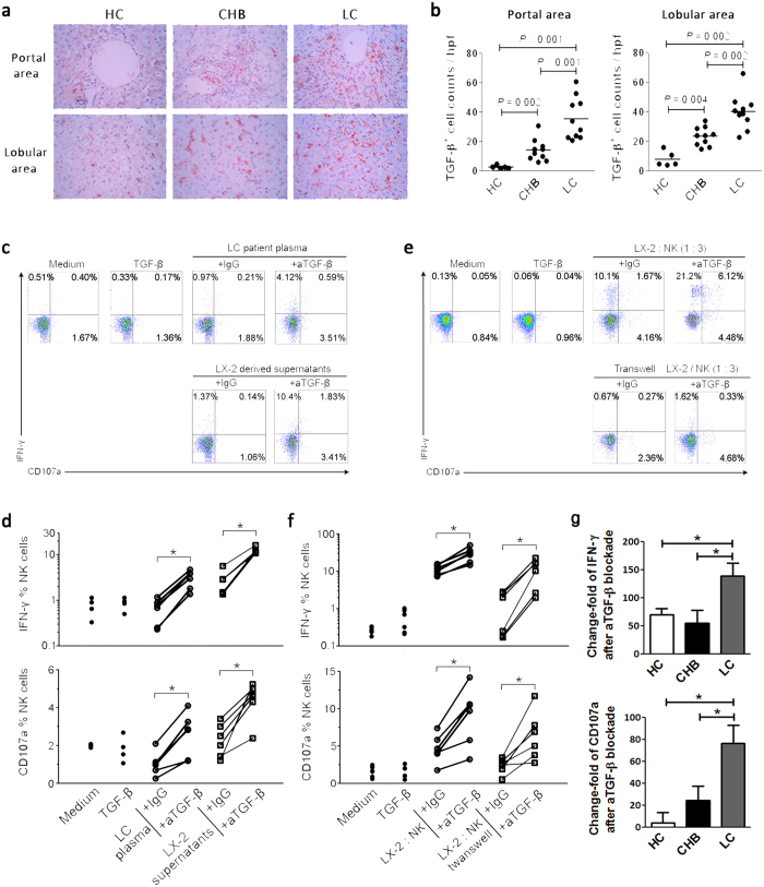
Figure 4. Impaired NK anti-fibrotic functions are dependent on TGF-β in LC patients.
(a) Representative IHC staining of hepatic TGF-β (red) in the portal and lobular areas from HC, CHB and LC subjects. (b) The quantitative analysis of TGF-β+ cells in the portal and lobular areas in the livers of the enrolled subjects. Each dot represents one individual. The horizontal lines indicate the mean values. P-values shown in the figures are based on two-tailed, unpaired Student’s t-test; hpf, high-power field. (c,e) Representative dot plots depicting IFN-γ and CD107a expression on peripheral NK cells from six HC subjects in response to plasma from LC patients or LX2 cell-derived supernatants (c), and LX2 cells co-culture or transwell assay (e). CD3−CD56+ NK cells were gated. Values in the quadrants represent the percentages of CD3−CD56+ NK cells that express IFN-γ and CD107a. (d,f) Blockade of TGF-β enhanced the production of CD107a and IFN-γ by NK cells significantly in LC plasma or LX2-derived supernatants (d), and LX2 cell culture or with transwell assay (f). (d,f) Each dot represents one individual. *P < 0.05, two-tailed, paired Student’s t-test. (g) Pool data indicated that anti-TGF-β treatment has more capacity to restore the production of CD107a and IFN-γ by NK cells from LC patients than that from HC and CHB individuals. Peripheral NK cells from HC (n = 4), CHB (n = 4) and LC (n = 8) individuals were co-cultured with LX-2 cells in vitro in the presence of IgG and anti-TGF-β antibodies. The change fold of CD107a or IFN-γ production by NK cells was calculate according to formula: (anti-TGF-β treatment - IgG) / IgG. *P < 0.05, two-tailed, unpaired Student’s t-test.

