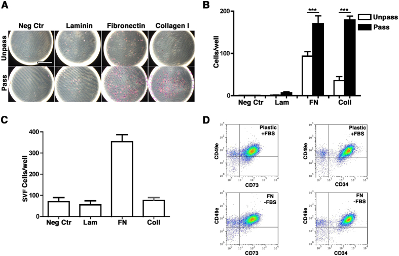
Figure 5.
(A) Representative images of the adhesion assay performed with Unpass- and Pass-ASC in the absence of any coated protein (Neg Ctr), or on wells coated with either laminin (Lam), fibronectin (FN) or collagen type I (Coll). Size bar = 400 μm; (B) Quantification of the number of adhered cells per well in the absence of any protein (Neg Ctr), or on wells coated with either laminin (Lam), fibronectin (FN) or collagen type I (Col I) for Unpass- (white bar) and Pass-ASC (black bar). ***p < 0.001 (ANOVA with Bonferroni multiple comparisons test); (C) Quantification of the number of freshly isolated SVF cells adhered per well; (D) Representative cytofluorimetry plots of SVF cells stained for CD49e and CD73 (left panels) or CD49e and CD34 (right panels) after overnight seeding on plastic in presence of FBS (upper panels) or on plastic coated with fibronectin in absence of FBS (lower panels).

