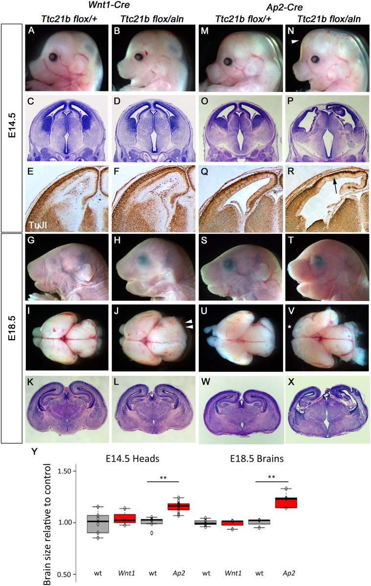
Fig 5. Deletion of Ttc21b from neural crest cells and surface ectoderm.
(A-L) Wnt1-Cre mediated deletion of Ttc21b does not lead to morphological (A,B,G,H,I,J), histological (C,D,K,L) or neural differentiation (E,F) phenotypes in the forebrain at E14.5 (A-F) or E18.5 (G-L). The midbrain is enlarged at E18.5 (double arrowheads in J). (M-X) Ap2-Cre; Ttc21bflox/aln embryos at E14.5 and E18.5 have an enlarged forebrain (arrowhead in N, V) with disrupted cortical architecture (P,X) and reduced numbers of differentiated neurons (R). Loss of olfactory bulbs is also noted at E18.5 (asterisk in V). All paired images are at the same magnification. (Y) Quantification for brain sizes. Center lines show the medians; box limits indicate the 25th and 75th percentiles as determined by R software; whiskers extend 1.5 times the interquartile range from the 25th and 75th percentiles, outliers are represented by dots; data points are plotted as open circles. n = 6, 3, 5, 12, 5, 3, 3, 5, respectively. (**: p <0.005).

