FIGURE 1:
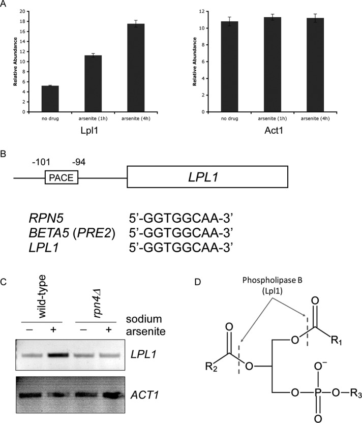
Lpl1 is regulated by Rpn4. (A) Relative protein abundance of Lpl1 at 0, 1, and 4 h after treatment with sodium arsenite (1 mM). Data were generated using a tandem mass tag-based mass spectrometry approach (Guerra-Moreno et al., 2015). Act1, a control protein, showed no change. Error bars represent SDs from triplicate cultures. In addition, differences between untreated and treated samples were statistically significant by Student's t test (p < 0.01) for Lpl1 but not Act1. (B) Schematic diagram of the LPL1 gene with its associated 5′-untranslated region. A classical PACE motif is present, with its location indicated relative to the start codon. This PACE motif is identical to that of other well-established Rpn4 targets, such as Rpn5 and Beta5 (Pre2). (C) Stress-inducible transcription of LPL1 in wild-type and rpn4Δ cells, as determined by RT-PCR. Treatment was with sodium arsenite (1 mM) for 1 h. ACT1 (bottom) serves as a control. (D) Schematic of a generic phospholipid with the cleavage sites indicated for type B phospholipases such as Lpl1. R1 and R2, fatty acyl groups; R3, polar head group (e.g., choline, ethanolamine, serine, inositol).

