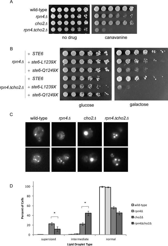
FIGURE 6:
Rpn4 recapitulates aspects of Lpl1 function. (A) Growth of wild-type, rpn4Δ, cho2Δ, and rpn4Δcho2Δ strains in the presence of canavanine (0.5 µg/ml). Cells were spotted in threefold serial dilutions and cultured at 30°C for 2–4 d. Note that a lower concentration of canavanine was used here than in Figure 4B. (B) Growth of rpn4Δ and rpn4Δcho2Δ strains expressing galactose-inducible wild-type STE6 or the misfolded mutant ste6-L1239X or ste6-Q1249X. Cells were spotted in threefold serial dilutions and cultured for 4 d at 33°C. The cho2Δ mutant alone did not show a phenotype under these conditions (unpublished data). (C) Representative images of lipid droplets, as visualized by fluorescence microscopy, for the indicated strains. cho2Δ cells show a relative accumulation of cells with a single very large (“supersized”) lipid droplet. Similar to cho2Δlpl1Δ, rpn4Δcho2Δ cells show a relative accumulation of cells with an intermediate phenotype (i.e., lipid droplets that are smaller but more numerous than those seen in cho2Δ). (D) Quantitation of normal, intermediate, and supersized lipid droplets. Six hundred cells were counted (in 100 cell groups) for each strain. Error bars represent SDs. Asterisk, statistically significant by Student's t test (p < 0.001). Note that all six strains represented in Figures 5A and 6C were analyzed as a group. Thus the values for wild-type and cho2Δ are the same in both.

