FIGURE 7:

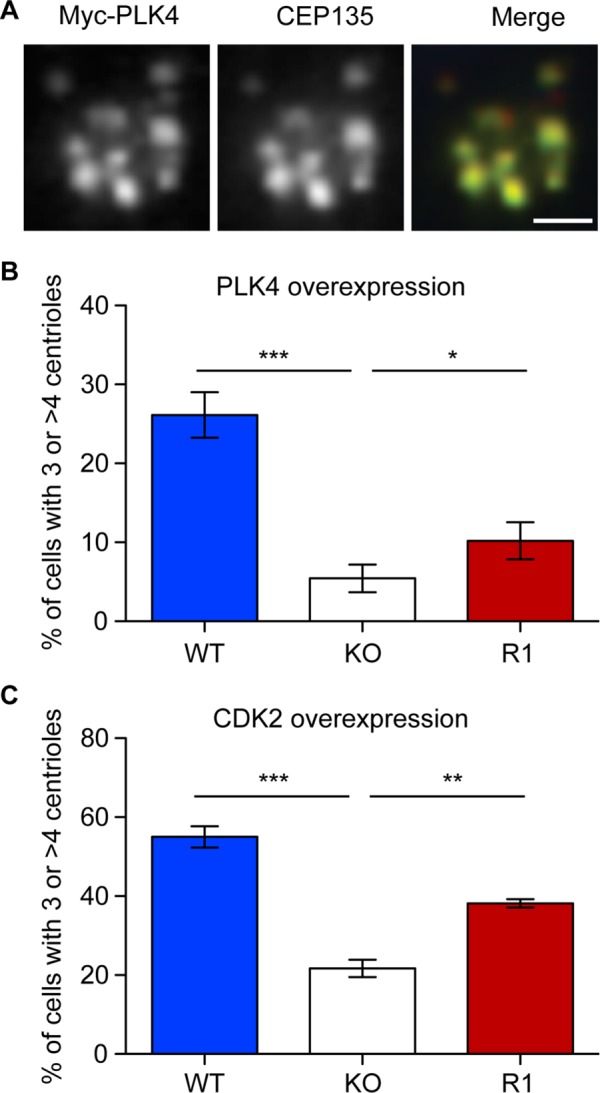
Reduced centrosome amplification induced by PLK4 and CDK2 overexpression in C-NAP1–deficient cells. (A) Immunofluorescence microscopy of a centriole rosette visualized with antibodies to myc (green) and CEP135 (red) 72 h after transfection of wild-type hTERT-RPE1 cells with a myc-PLK4 overexpression construct. Scale bar, 2 μm. (B, C) Centrosome quantitation in cells of the indicated genotype 72 h after transfection with constructs encoding (B) myc-PLK4 or (C) HA-CDK2. Centrioles were scored by staining with antibodies against CEP135 or centrin and transfected cells identified using antibodies to myc or CDK2. Bar graphs indicate mean ± SD of three separate experiments in which at least 50 transfected cells were counted. ***p < 0.001, **p < 0.01, and *p < 0.05 by unpaired t test.
