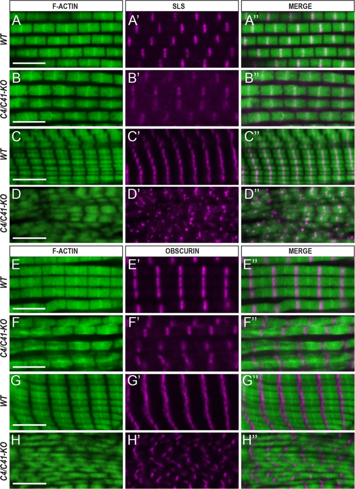
FIGURE 5:
TpnC is required for myofibril assembly. Confocal images of cryosectioned IFM and TDT muscles from WT and TpnC41C∂#2 TpnC4∂#25 double-knockout mutants stained for F-actin, Sallimus (Sls), and Obscurin. (A-A″, B–B″, C–C″, D–D″) Staining with phalloidin (green) and anti-Sls antibody (magenta). (A-A″, B–B″) IFM. (C–C″, D–D″) TDT myofibrils. (E–E″, F–F″, G–G″, H–H″) Immunostaining with phalloidin (green) and anti-Obscurin antibody (magenta). (E–E″, F–F″) IFM. (G–G″, H–H″) Images of TDT. Scale bar, 5 μm. All images represent longitudinal sections of the muscles. Note that myofibril organization is disrupted in the mutants compared with controls.

