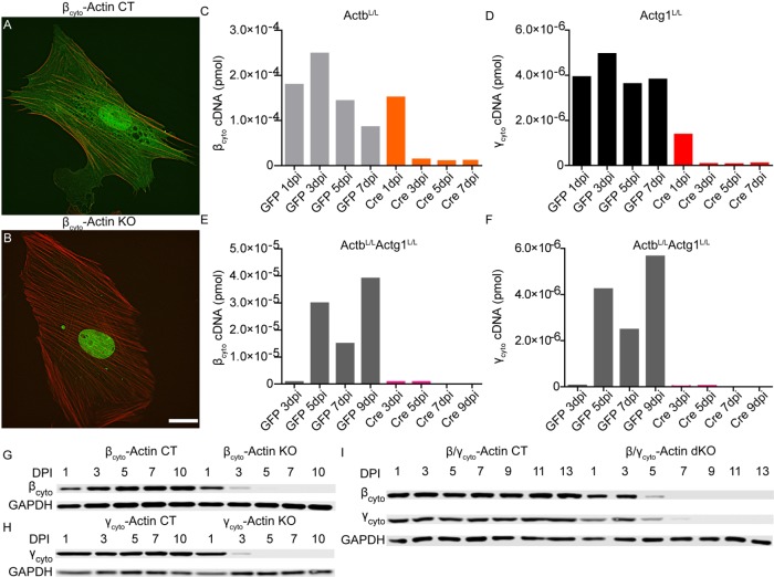
FIGURE 1:
Adenoviral Cre efficiently ablated βcyto- and γcyto-actin in primary MEFs. (A, B) Representative images of βcyto-actin CT and βcyto-actin KO cells at 7 dpi. Scale bar, 20 µm. (C–F) Representative qRT-PCR analysis of βcyto- and γcyto-actin transcript amount (picomoles) in ActbL/L, Actg1L/L, and ActbL/L Actg1L/L MEFs. (G–I) Representative relative Western blot analysis of MEF lysates probed with βcyto-actin and γcyto-actin antibodies; GAPDH served as loading control.

