
FIGURE 10:
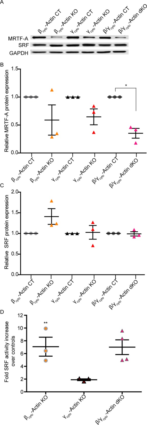
SRF activity but not protein was up-regulated in βcyto-actin–ablated MEFs. (A) Representative Western blot analysis of CT and KO MEFs blotted with MRTF-A and SRF; GAPDH served as loading control. (B, C) Relative protein expression normalized to GAPDH and relative to the paired embryo control (n = 3). (D) Calculated fold increase in SRF activity, via luciferase assay, in KO over CT MEFs (n = 3). *p < 0.05, **p < 0.01. One-sample t test; error bars are SEM.