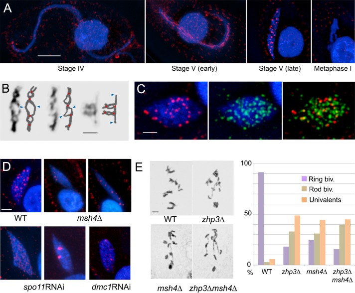
FIGURE 3:
Zhp3 localization and mutant phenotypes. (A) Zhp3 localization at different meiotic stages in the wild type (background staining outside nuclei comes from basal bodies of cilia). (B) Individual diplotene bivalents with schematic interpretations. Possible CO sites and centromere regions are indicated by red dots and blue arrowheads, respectively. (C) BrdU (incorporated during recombinational repair; green) and Zhp3 (red) coappear at late stage V. (D) Zhp3 foci in wild-type (WT), msh4Δ, spo11RNAi, and dmc1RNAi cells. (E) Quantification of ring bivalents, rod bivalents, and univalent pairs in WT, zhp3Δ and msh4Δ single mutants, and zhp3Δmsh4Δ double mutant. Bars, in 10 µm (A–D), 2 µm (E).

