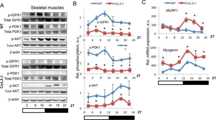
FIGURE 3:
IGF signaling is reduced in skeletal muscle of Cry1, 2−/− mice. (A) Representative Western blotting (pooled extracts from three mice per time point) of phospho–IGF-1R on Y1137/1138, PDK1 on S241, and AKT on S473 in skeletal muscle of WT (blue diamonds) and Cry1, 2−/− (red squares) male mice. (B) Quantification of phospho–IGF-1R on Y1137/1138, PDK1 on S241, and AKT on S473 in skeletal muscle of WT (blue diamonds) and Cry1, 2−/− (red squares) male mice (N = 3 per time point). (C) mRNA expression of FOXO transcriptional targets in skeletal muscle of WT (blue diamonds) and Cry1, 2−/− (red squares) male mice (N = 3 per time point). *Statistically significant difference between genotypes (p < 0.05). Light was on at ZT0 and off at ZT12.

