
Figure 3. The crystal structure of the uncomplexed CyRPA.
(A) Orthogonal view of the ribbon representation of CyRPA, colored in rainbow fashion from the N-terminus (blue) through to the C- terminus (red). (B) Amino acid sequence and the secondary structure of CyRPA, showing the location of the 24 canonical strands of the six-bladed β-propeller [labelled ‘βmsn’ where the index n denotes the strand and the index m the sheet (n = 1,..,4; m = 1,..,6)]. (C) Assembly of the 222 pseudo-symmetric tetramer of CyRPA within the crystallographic asymmetric unit, showing the formation of the extended 5–5 and 6–6 sheets (orange and red, respectively). Each monomer of CyRPA in the asymmetric unit is labelled a, b, c, d.

Figure 3—figure supplement 1. Comparison of CyRPA with C.

perfringens NanI sialidase. Clustal Omega sequence alignment of P. falciparum CyRPA (without signal peptide) and the catalytic domain of Clostridium perfringens NanI sialidase (Newstead et al., 2008). These sequences share 16% identity and 33% similarity. Conserved sialidase features include Asp box motifs (red boxes), catalytic acid/base residue (green), catalytic nucleophile residue (yellow) and the acidic residue that modulates the nucleophile’s pKa (blue).
Figure 3—figure supplement 2. Recombinant CyRPA does not have sialidase activity.
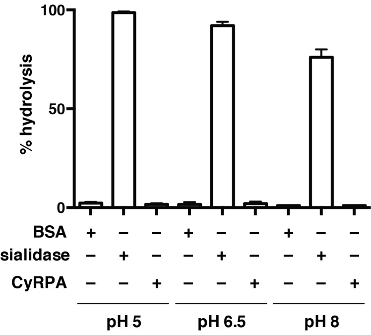
Hydrolysis of 4-methylumbelliferyl N-acetyl-α-D-neuraminic acid (4MU-NeuNAc) in the presence of bacterial sialidase (neuraminidase) from Arthrobacter ureafaciens, CyRPA or BSA at different pH values. These values, obtained in triplicate, were normalized with respect to the fluorescence measured for 10 μM 4-methylumbelliferone in glycine buffer (0.9 M, pH 10) to determine the completeness of each reaction.