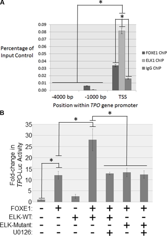
Figure 5. FOXE1 and ELK1 interact with the TPO gene promoter.
(A) Detection of FOXE1 and ELK1 binding at the TPO gene promoter by ChIP. Sheared formaldehyde-fixed chromatin was isolated from thyroid tissue and then immunoprecipitated with monoclonal antibodies raised against human FOXE1 and ELK1 proteins. ChIP DNA was amplified by real-time qPCR using primers specific for the proximal TPO promoter and two negative control regions located 1000- and 4000-bp upstream of the TSS. Enrichment of transcription factor binding was calculated as a percentage of the input DNA control. Values are the mean average and SD of three independent experiments. Significant enrichments over IgG controls are highlighted (*p < 0.01, Student’s t-test). (B) Characterizing FOXE1-ELK1 mediated regulation of the TPO gene promoter. NThy cells were transiently transfected with TPO-luc and different combinations of FOXE1, ELK1, mutant ELK1 or empty expression plasmids. Twenty-four hours post-transfection the cells were treated for a further 24 hrs with 10 μM U0126 or vehicle, prior to whole cells lysates being harvested for luciferase reporter assays. Luciferase results are the the mean (± SD) of three experiments, both performed in triplicate, expressed as fold increase in luciferase activity relative to empty vector transfected cells. Significant changes are highlighted (*p < 0.05, Student’s t-test).

