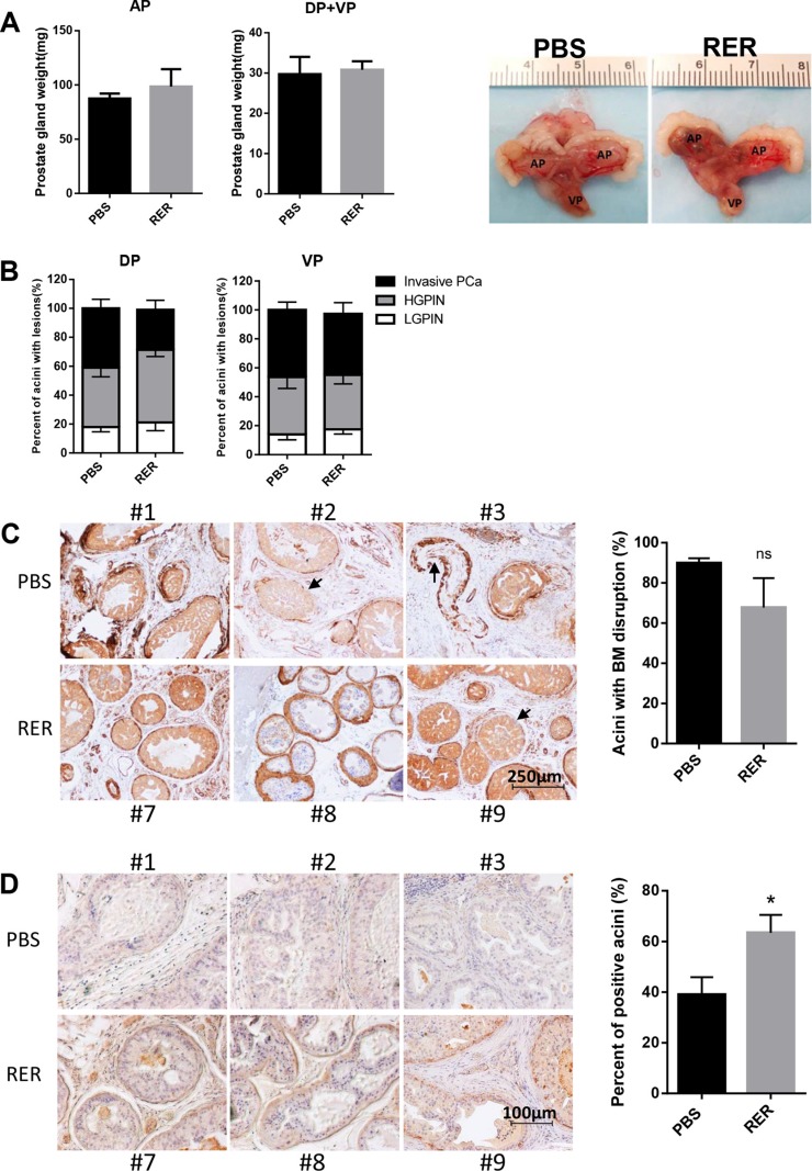
Figure 6. RER treatment moderately blocked early stage prostate cancer progression in vivo.
(A) Each prostate gland was isolated from mice and weight was recorded. Bar plots show the weight of prostate glands in PBS and RER group. Representative pictures of prostate glands from PBS and RER-treated mice are presented. (B) Percent of normal/low grade PIN lesion, high grade PIN lesion, and invasive adenocarcinoma in each prostate lobe was obtained by dividing the number of acini with the specific lesion by the total number of acini observed in one tissue section. (C) IHC analyses of α-SMA in DP lobes showed that RER treatment group had a moderately less breakdown of a continuous layer of basement membrane (BM) around each acinus. (D) IHC analysis of laminin in DP lobes showed that more acini in RER group had positive laminin staining than in Control group. All plotted data represent mean±s.e.m. for six mice. “n.s.” denotes not significantly different and “*” denotes significantly different at P < 0.05.

