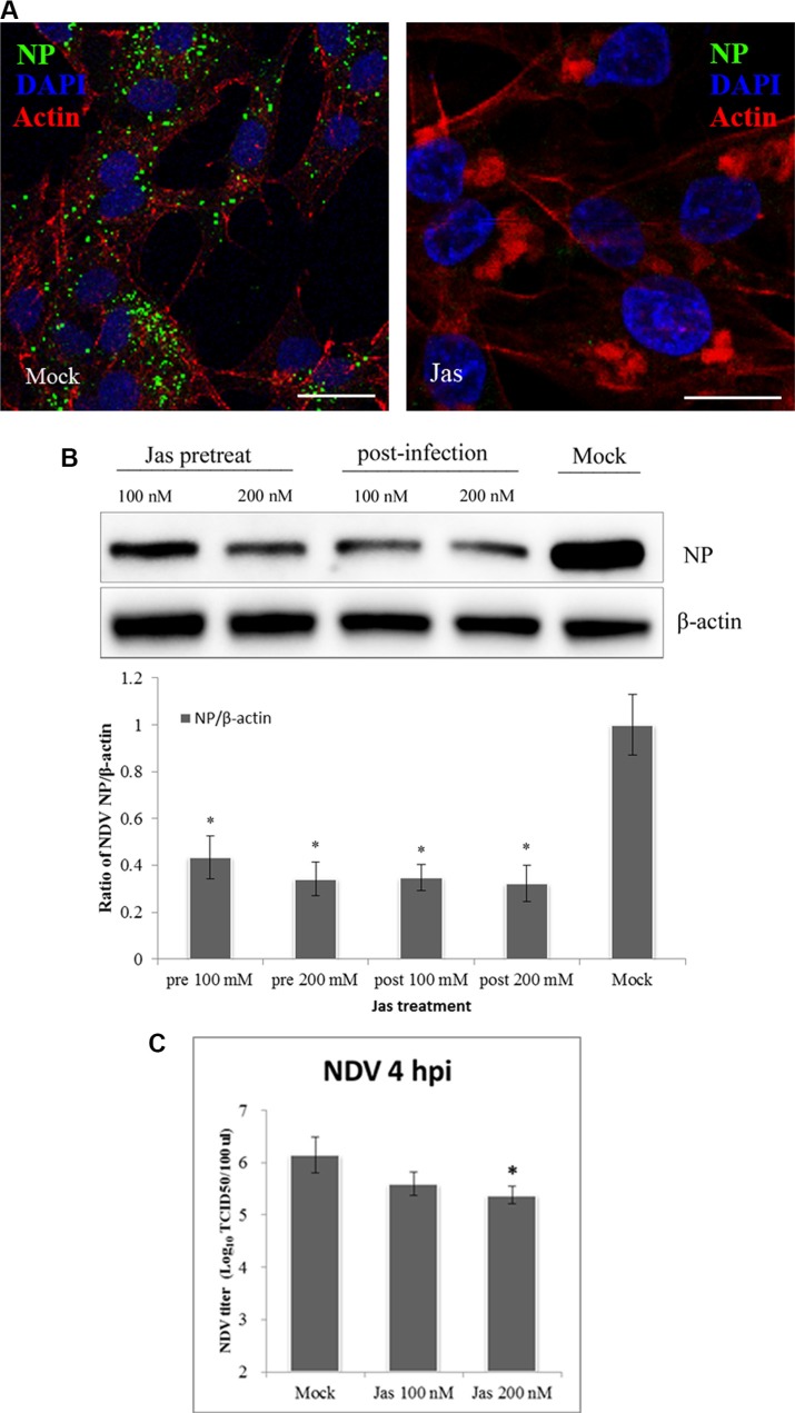
Figure 4. Actin rearrangements during NDV internalization.
(A) Jasplakinolide inhibition of NDV entry into DF-1 cells. Pretreated cells (200 nM jasplakinolide) were infected with 5 MOI NDV for 4 h at 37°C and observed using CLSM: Actin filaments (red), NP (green), and cell nucleus (blue). (B) Western blotting showing jasplakinolide inhibition of NDV entry as indicated by NP levels. The ratio of NP/b-actin is shown. (C) The NDV titers of the supernatant of Mock- and jasplakinolide-treated cells were determined using TCID50 assays. The results are presented as the mean ± SD, *P < 0.05, Scale bar = 20 μm.

