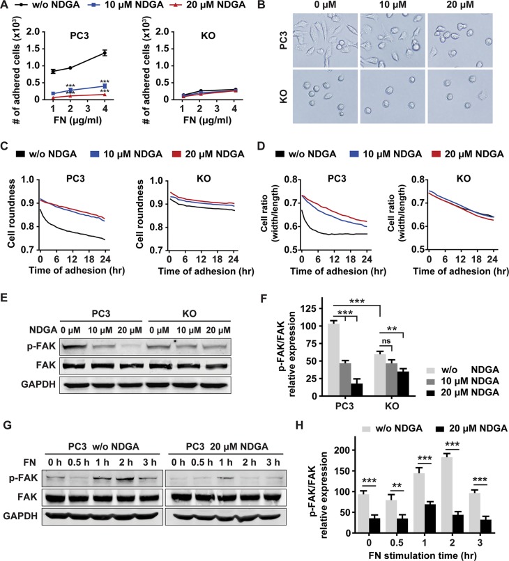
Figure 4. NDGA inhibits tumor cell-fibronectin adhesion and FAK activation.
(A) PC3 and KO cells were pretreated with 0, 10 or 20 μM NDGA for 24 hours and then seeded into FN-coated wells allowing for 30 minutes adhesion. Numbers of adhered cells of each group are counted. Data show mean ± S.E (n = 3). ***p < 0.001. (B) Pretreated cells were seeded into FN-coated wells and representative images of cell morphology after 24 hours adhesion were shown. (C, D) Morphological analysis of cells during adhesion progress. Cell adhesion to FN matrix was monitored in live imaging system for 24 hours. Cell roundness (C) and cell width/length ratio (D) of both PC3 and KO cells are shown. (E, F) Western blot analysis of p-FAK (Tyr379) and total FAK expression after NDGA treatment for 24 hours. All data are normalized to the control group of PC3 cells and results present mean ± S.E (n = 3). **p < 0.01, ***p < 0.001. (G, H) Western blot analysis of p-FAK (Tyr379) and total FAK expression levels under short term stimulation of FN in PC3 cells with or without 20 μM NDGA pretreatment. All data are normalized to the PC3 cells without FN stimulation. Data present mean ± S.E (n = 3). **p < 0.01, ***p < 0.001.

