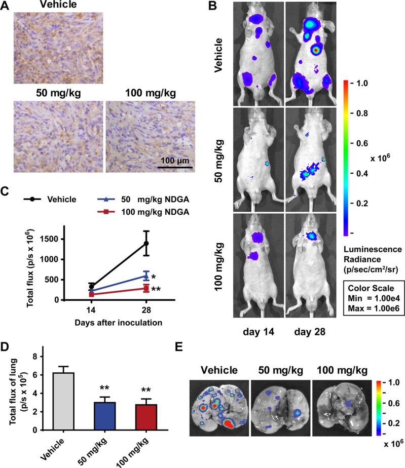
Figure 6. NDGA inhibits PC3 xenografts growth and metastasis.
(A) Immunohistochemistry staining of NRP1 in the tumor tissues of subcutaneous xenograft model. Scale bar: 100 μm. (B) Representative bioluminescence images showing the metastatic sites in intravenous injection model. Mice of three groups (vehicle, 50 mg/kg NDGA, 100 mg/kg NDGA) were monitored after 14 days and 28 days drug treatments. (C) Quantification of bioluminescence signal within the region of interest at day 14 and day 28. Data show mean ± S.E (n = 5). *p < 0.05, **p < 0.01. (D) Quantification of bioluminescence signal of lungs at day 28. Data show mean ± S.E (n = 5). **p < 0.01. (E) Representative bioluminescence images of lungs with metastatic sites.

