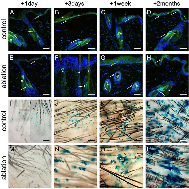
Figure 3. Lgr6+ stem cells and their progeny in haired mice after UV overexposure.

Anti-EGFP staining was used to localize the Lgr6+ stem cells A-H. and whole mounts were stained for LacZ+ progeny I-P. at different time points. We observed some autofluorescence in hairs. The control mice (A-D) showed clear EGFP+ cells in the IFE and in the isthmus (A-D, see arrows). Lgr6 progeny built up in the HFs and islands in the IFE (I-L). One day after UV overexposure we still found some EGFP expression in the IFE (E, in apoptotic basal layer), that expression was lost after 3 days (F, with inflammatory infiltrate in papillary dermis) and reappeared sparsely after 1 week (G). We found EGFP expression in the isthmus at all time points (E-H). One day after UV overexposure we only observed some LacZ+ cells in the HFs and not at all in the IFE (I). From three days after UV overexposure the Lgr6 progeny was found in the IFE, more associated with HFs remnants (N-P) compared to the controls. HF orifices indicated by dotted lines (O+P). Scale bars = 50 μm. *= background autofluorescence in sebaceous glands and from hairs.
