
Figure 7. Lgr6 progeny in inter-tumoral skin and skin tumors of haired mice.
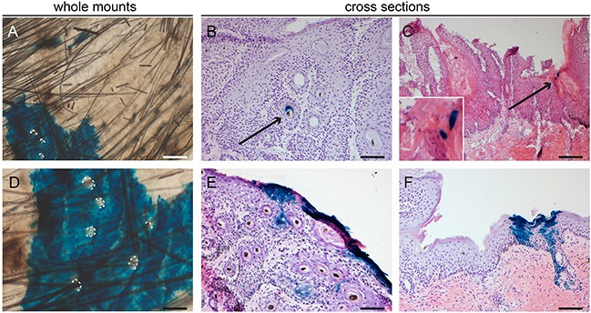
Samples subjected to chemical carcinogenesis were stained for LacZ+ cells. Whole mount inter-tumoral skin after chemical carcinogenesis A+D. showed large islands of Lgr6 progeny in the IFE when lineage tracing was induced at the start of the experiment. Hair follicle orifices contoured in A+D of hair follicles that did not stain. Only after early lineage tracing some tumors showed incidental staining in engulfed hair follicle-like structures B. We found more abundant staining in uninvolved skin adjacent to the tumors, notice features of proliferating units F. When lineage tracing was induced when tumors were formed we found some incidental staining in differentiated cells (C and insert). E. shows the only tumor (out of 43) in haired mice subjected to chemical carcinogenesis that showed some staining at the outer rim of the tumor with early tracing. Scale bars = 100 μm (B-F) and 200 μm (A).