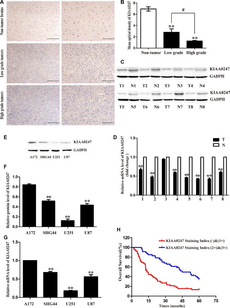
Figure 1. Low KIAA0247 expression predicted a worse prognosis for patients with glioma.
(A). Representative images of KIAA0247 immunostaining in non-tumor brain tissues, low grade-glioma tissues (WHO I-II) and high-grade glioma tissues (WHO III-IV). Low power (20×) scale bars: 100 μm, high power (40×) scale bars: 50 μm. (B) Statistical quantification of the mean optical density in non-tumor brain tissues, low-grade glioma tissues, and high-grade glioma tissues (**P < 0.01 versus non-tumor brain tissues; #P < 0.05 between the high-grade group and low-grade group). (C) Western blot shows KIAA0247 expression was lower in 8 pairs of predominately glioma tissues (T) compared to paired adjacent non-tumor tissues (N). (D) qRT-PCR analysis of KIAA0247 mRNA in 8 glioma tissues and paired adjacent non-tumor tissues (**P < 0.01 versus paired adjacent non-tumor tissues). (E, F) Western blot analysis of endogenous expression of KIAA0247 in four glioma cell lines (**P < 0.01 versus A172). (G) qRT-PCR analysis of endogenous KIAA0247 expression in four glioma cell lines (**P < 0.01 versus A172). (H) Kaplan-Meier analysis of overall survival based on KIAA0247 expression in 112 patients with glioma (P < 0.01).

