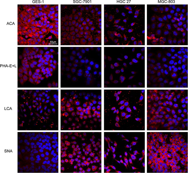
Figure 2. Expression of glycans in the four cell lines evaluated by lectin staining.

GES-1, SGC-7901, HGC-27, and MGC-803cells were stained with Cy3-labeled lectins (ACA, PHA-E+L, LCA, SNA). Signals in merge images of Cy3-conjugated lectins and DAPI staining of nuclei are shown. Objective magnification: 60x.
