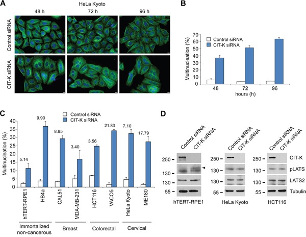
Figure 3. CIT-K is essential for the completion of cytokinesis in a panel of cancer cell lines.

A. HeLa Kyoto cells were plated on coverslips and treated with a siRNA against either a random sequence (control) or CIT-K for 48, 72 and 96 hours (h), where cells were fixed and stained to detect tubulin (green) and DNA (blue). Bars, 10 μm. B. Quantification of multinucleation increase from the experiment shown in A. At least 600 cells were counted for analysis in each experiment, n=3, error bars represent SEM. C. Quantification of the percentage of multinucleated cells (of cell lines listed) that were treated with either control or CIT-K siRNA for 48 h. Cells were fixed and stained to detect tubulin (green), phalloidin (red) or DNA (blue). The fold difference between control and CIT-K siRNA-treated cells is listed above each cell line. At least 600 cells were counted for analysis in each experiment, n=3, error bars represent SEM. D. hTERT-RPE1 (left panel), HeLa Kyoto (middle panel) and HCT116 (right panel) cells were treated with either control or CIT-K siRNA for 48 h, harvested and protein extracts analyzed by Western blot to detect CIT-K, phosphorylated LATS (pLATS), LATS2 and tubulin. The numbers on the left indicate the sizes in kilodaltons of the molecular mass marker.
