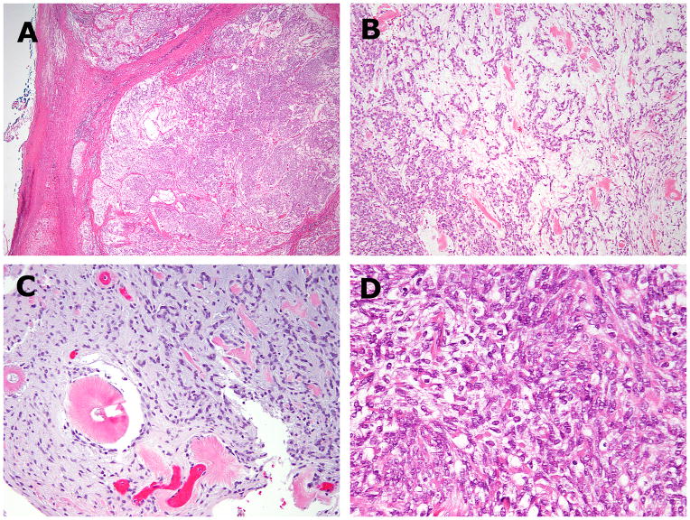
Figure 2. Histologic findings of EWSR1-CREM positive index case 1.
(A,B) The pelvic tumor showed circumscribed borders and a lobulated growth, composed of alternating hypo and hyper-cellular areas. (C) The hypocellular myxoid areas showed uniform round to ovoid cells arranged in cord-like or reticular patterns. Scattered amianthoid fibers were seen in both the primary (B) and recurrent tumors (C). (D) Cellular areas showed densely packed cells with indistinct cell border and palely eosinophilic cytoplasm.

