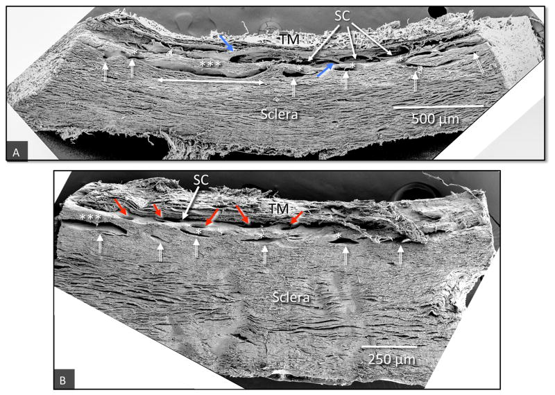
Fig. 9.
Scanning electron microscopy of tilted frontal sections providing images of regions of the limbal circumference that include the trabecular meshwork, (TM), Schlemm’s canal (SC) and a series of intrascleral collector channels ISCC (white double bar arrows) in the deep scleral plexus. (A) The ISCC are distributed in a relatively narrow region close to SC. Rather than being round the ISCC have long dimensions’ parallel to and a short dimension perpendicular to SC. The ISCC configuration results in thin septa (*), attached at their ends to the sclera that are at times have a length many times their height (***). Blue arrows identify cylindrical structures crossing SC that connect the trabecular meshwork with regions of SC external wall near CCE. (B) The collector channel entrances (CCE) are depicted by red arrows (non-human primate, M. fascicularis).

