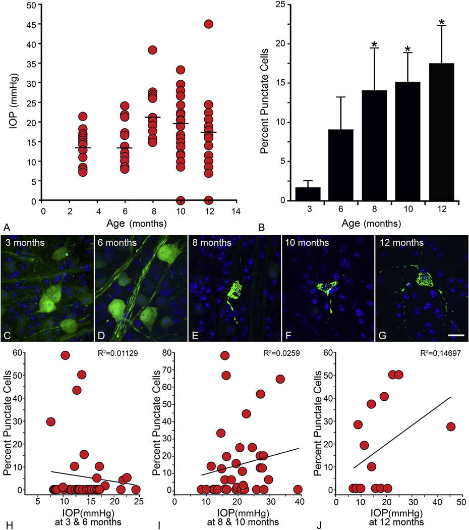
Fig. 5.
DBA/2J glaucoma is accompanied by the activation of GFP-BAX. In this experiment, DBA/2J mice were housed to different ages and then processed to determine the localization of GFP-BAX. One month prior to euthanasia, mice were injected with the AAV2-GFP-BAX virus. (A) Intraocular pressure (IOP) measurements of eyes at time of euthanasia. In this experiment, 19 eyes were evaluated at 3 months, 20 at 6 months, 17 at 8 months, 22 at 10 months, and 16 at 12 months. As the mice get older, IOP levels generally increase (mean IOP denoted by the straight line in each scatter plot). (B) Graph showing the change in GFP-BAX localization as a function of age. Young (3 month) mice show almost no punctate cells (punctate localization was detected in only 4 of 18 eyes). More punctate cells are detected at 6 months, when disease-related changes are first evident. This was highly variable, however, and not significantly greater than the 3 month cohort. A significant increase in the punctate localization was detected at all ages between 8 and 12 months (*P < 0.05). Overall, this pattern reflects other glaucoma-related pathology described in this strain (Libby et al., 2005b; Schlamp et al., 2006). The total number of cells counted were: 3 months–713; 6 months–560; 8 months–588; 10 months–254; 12 months–221. (C–G) Images of labeled cells showing the representative localization pattern of GFP-BAX as a function of age. Size bar = 10 µm (H–J) To assess the correlation between IOP at euthanasia and GFP-BAX localization, the data was stratified into younger (3 and 6 month old, H), mid-aged (8 and 10 months, I), and old mice (12 months, J) and the percent punctate labeled cells for each eye was graphed as a function of IOP at death. The correlation coefficients for each dataset show no significant association between the two metrics.

