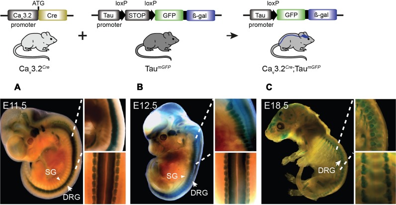
FIGURE 3.
Cre activation in Cav3.2Cre; TaumGFP mice in development. β-gal expression in the nervous system of Cav3.2Cre;TaumGFP mice as shown by the blue staining. Embryos at stages (A) E11.5, (B) E12.5, and (C) E18.5 of development. Note the stronger β-gal staining in the spinal cord and DRGs in early stages compared to the brain. The staining observed in bones in the E18.5 embryos was also observed in negative controls. Zoom in panels: lateral and dorsal views of the spinal cord. DRG, dorsal root ganglia indicated with arrows; SG, sympathetic ganglia indicated with arrowheads.

