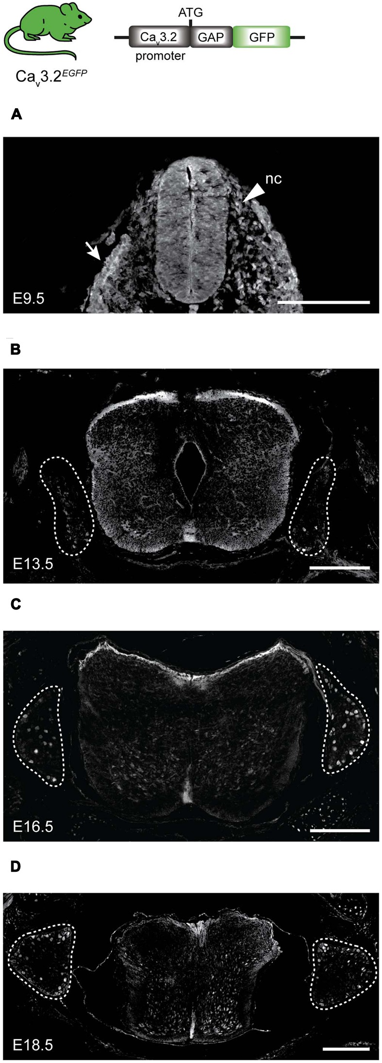
FIGURE 5.

EGFP expression during development in spinal cord and DRG of Cav3.2EGFP knock-in mice. (A) At E9.5, EGFP is expressed in some cells of the neuronal crest (nc) indicated by an arrow head, in the epithelium indicated by an arrow, and in the spinal cord. (B) At E13.5, a few cells of the DRG are EGFP+, while the staining at the dorsal side of the spinal cord is stronger. (C) At E16.5 the number of EGFP+ cells increases in the DRG, but staining in the spinal cord is reduced. (D) Finally at E18.5, EGFP expression in the DRG appears constant while the positive staining has almost disappeared in the spinal cord. Dashed lines outline the DRGs. Scale bars: 200 μm.
