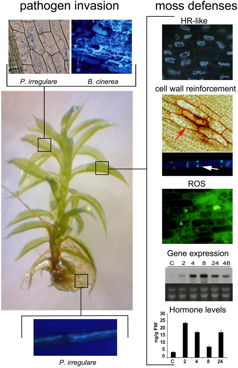
FIGURE 1.
Pathogen invasion and P. patens defense responses. P. patens has one layer of cells in most tissues facilitating microscopic analysis of pathogen colonization processes and moss defense mechanisms (Ponce de León, 2011; Overdijk et al., 2016). Leaves and rhizoids are colonized by different pathogens, which are easily visualized by different staining techniques (leaf; trypan blue and solophenyl flavine, rhizoid: solophenyl flavine) (Ponce de León and Montesano, 2013). Representative pictures of the oomycete P. irregulare and the fungus B. cinerea pathogen-infected tissues are shown. Plant defenses are activated after pathogen infection, including a HR-like response, cell wall fortification and chloroplast reorientation (red arrow), ROS accumulation, defense gene expression and increase in hormone levels (Ponce de León and Montesano, 2013). Representative pictures of defense responses are shown; HR-response, cell wall fortification (phenolic compounds accumulation, evidenced by stained cell walls) and ROS production in B. cinerea-infected leaves, callose deposition in P.c. carotovorum elictor treated protonemal cells (white arrow), and expression pattern of the defense gene Ppalpha-DOX and auxin levels in response to elicitors of P.c. carotovorum at different hours after treatment. The HR-like response was visualized by autofluorescent compounds, cell wall fortification by safranin-O staining, callose deposition by methyl blue staining, and intracellular ROS with 2′,7′-dichlorodihydrofluorescein diacetate. “Graph showing auxin levels as originally published in Alvarez et al. (2016).”

