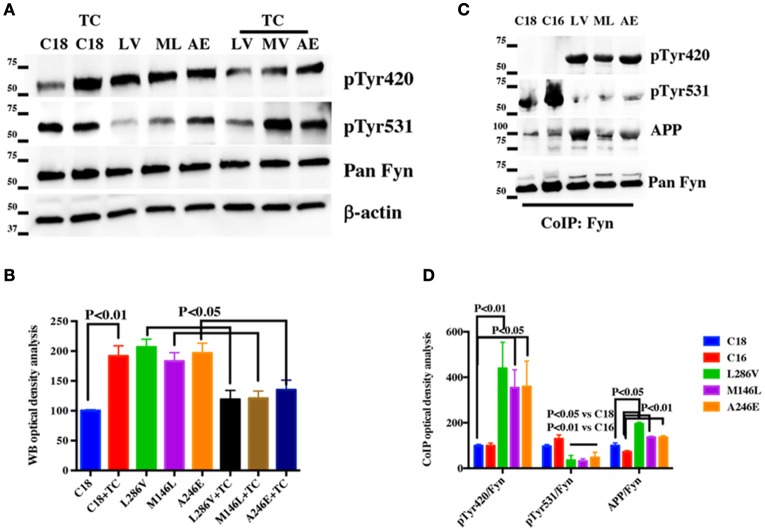
Figure 6.
The Tyr kinase Fyn binds APP in AD neurons: (A) WB analysis performed on total lysate from control (C18) and AD neurons before or after TC2153 (TC) exposure. Samples were probed with Src pTyr416 and Src pTyr527 antibodies used to analyse Fyn pTyr420 and Fyn pTyr531 levels and were normalized to the corresponding total Fyn levels and expressed as % of control. β-actin was used as loading control (B). (C) IP analysis of controls (C18 and C16) and AD neurons (LV, ML, and AE). Samples were immunoprecipitated with rabbit anti-pan Fyn antibody and analyzed using rabbit anti-Src pTyr416 and rabbit anti- Src pTyr527 antibodies (used to detect Fyn pTyr420 and pTyr531 levels; Xu et al., 2015) or mouse anti-APP (clone Y188). Data were normalized to the corresponding input Fyn levels, and expressed as % of the correspondent C18 values (D). Densitometric analysis of (A,C) is reported in (B,D). Statistically significant differences were calculated by one-way ANOVA and Tukey's post hoc test.

