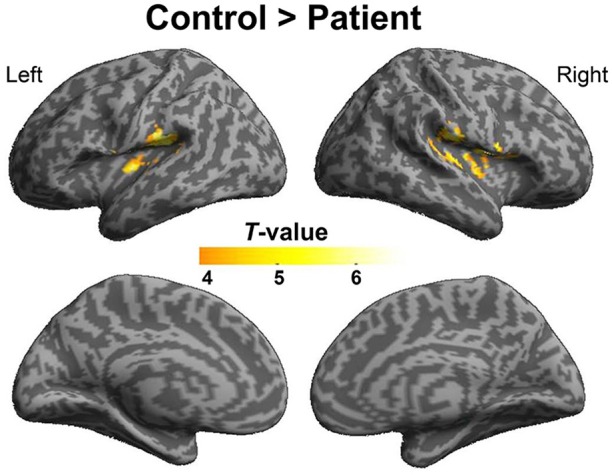
Figure 7.

Difference in brain regions exhibiting differential psychophysical interaction with left posterior inferior temporal gyrus associated with “VSP vs. VNSP” between healthy participants and people with schizophrenia. The activation maps were thresholded at p < 0.05 (voxel-wise FDR corrected, T > 4.86) and overlaid on a template brain surface of inflated cortex from SPM8. VSP, visual speech priming; VNSP, visual non-speech priming.
