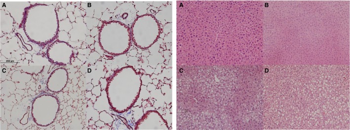
Figure 4.

(A) Lung sections from diet cohorts do not show overt inflammation or gross structural changes. Representative images of Masson's trichrome stained lungs (40X). (B) Liver sections stained hematoxylin and eosin stained liver (20X) reveal vacuolization only in offspring cohorts actively consuming HFD. All images are of offspring at 10 weeks of age. Diet cohort legend: NF is normal fat content diet, HF is high fat content diet. The first abbreviation indicates the maternal diet, the second following the hyphen indicates the postnatal diet. A. NF‐NF, B. HF‐NF, C. NF‐HF and D. HF‐HF.
