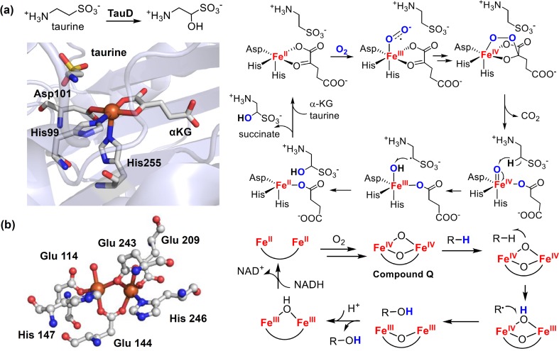
Fig. 8.
a Active site structure of a typical αKG dependent non-heme iron(II) dioxygenase, TauD, (PDB: 1OS7) and the mechanism of taurine hydroxylation catalyzed by TauD. b Active site structure of a representative non-heme diiron hydroxylase, soluble methane monooxygenase (sMMO) in reduced state (PDB: 1FYZ) and the typical mechanism of C–H hydroxylation catalyzed by non-heme diiron hydroxylase. Colors: iron (orange), oxygen (red), nitrogen (blue), sulfur (yellow)

