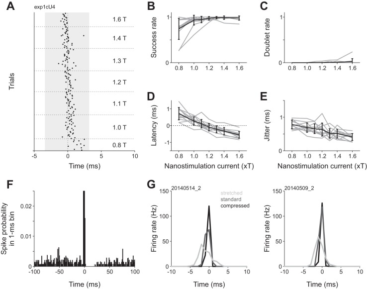
Fig. 2.
Current amplitude dependence of spike induction by kurzpuls nanostimulation. A: spike raster for an example neuron stimulated with kurzpulses of different amplitudes. Stimuli were presented in pseudorandom sequence (repetition rate 0.4 Hz), but are sorted for display purposes. Current amplitudes are expressed as a factor of threshold current T (8.5 nA for this neuron). Shading indicates the extent of the central positive peak of the kurzpuls (duration 7.0 ms, see Fig. 1B). B: fraction of trials in which kurzpuls nanostimulation evoked one or more spikes inside a 7-ms analysis window centered on the kurzpuls peak (“success rate”) as a function of nanostimulation current. Shaded lines: individual neurons, solid line: success rate averaged over n = 12 cells. Error bars denote standard deviation. C: fraction of trials in which kurzpuls nanostimulation evoked two or more spikes (“doublet rate”). Conventions in C–E are the same as in B. D: spike onset latency (relative to kurzpuls peak); positive values indicate that spikes lagged the pulse peak. E: spike time jitter, computed as the standard deviation of first spikes evoked by the kurzpulses. F: average PSTH of 10 neurons centered on kurzpuls peaks (1.3 T, repetition rate 0.4 Hz). Bin size, 1 ms. Ordinate was truncated (PSTH extends up to a probability of ~0.7). G: PSTHs from two cells exposed to modified kurzpulses of different widths. Kurzpuls waveforms were modified by changing the output sampling rate (standard 10 kHz), resulting in either compressed (20 kHz) or stretched (5 kHz) waveforms. Single pulses were delivered at 1.3 T with a repetition rate of 0.4 Hz. Thresholds were determined separately for each waveform (T values, cell 1: 5.5, 7.0, 9.5 nA; cell 2: 5.0, 5.0, 9.5 nA for stretched, standard, and compressed kurzpulses, respectively). Analysis windows extended from −3.5 to +3.5 ms relative to the kurzpuls peaks. Success rates were equally high for all waveforms (>0.97), whereas doublet rates were 0 except for the stretched kurzpuls presented to cell 1 (0.13). Spike time jitter was 1.2 (1.0), 0.6 (0.5), and 0.2 (0.3) ms for stretched, standard, and compressed kurzpulses for the cell on the left and right, respectively. Data on the left are from the neuron depicted in Fig. 1.

