Figure 1.
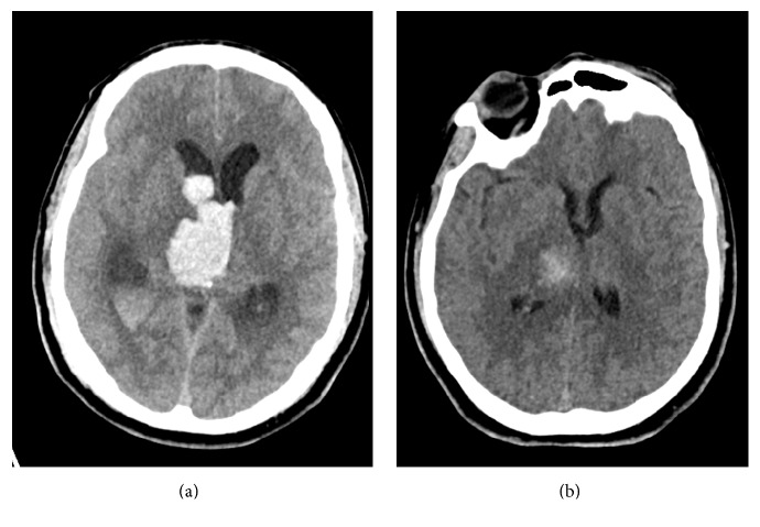
Computed tomography (CT) of the head. (a) CT head on admission showing a right thalamic hemorrhage with intraventricular extension. Note the obstructive hydrocephalus. (b) CT head on hospital day 9 showing evolution of the right thalamic hemorrhage and resolution of the obstructive hydrocephalus with external ventricular drainage (not shown) and after intraventricular tissue plasminogen activator (tPA).

