Figure 1.

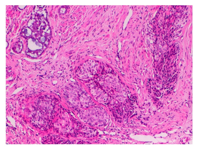
Photomicrograph of biopsy specimen of Case 1, showing the squamous cell carcinoma component (middle) and the adenoid cystic carcinoma component (upper left) (Hematoxylin and Eosin ×100).

Photomicrograph of biopsy specimen of Case 1, showing the squamous cell carcinoma component (middle) and the adenoid cystic carcinoma component (upper left) (Hematoxylin and Eosin ×100).