Figure 1.
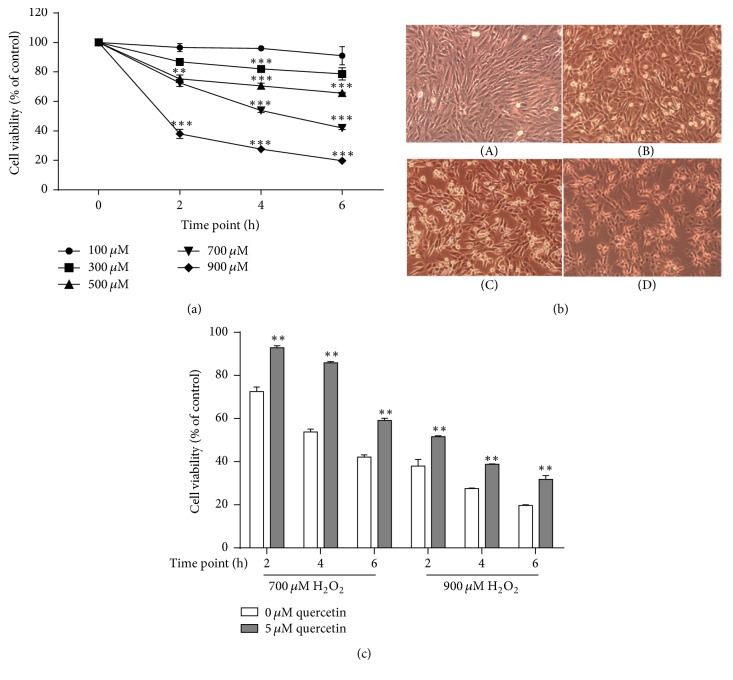
Oxidative damage model establishment in H9c2 cardiomyocytes. (a) H9c2 cells were treated with various concentrations of H2O2 (100 μM, 300 μM, 500 μM, 700 μM, and 900 μM) for different time periods (2, 4, and 6 h). (b) H9c2 cell morphology from 700 μM H2O2 stimulation for 0 h (A), 2 h (B), 4 h (C), and 6 h (D). (c) Comparisons of cell survival rates between quercetin treatment group and oxidative injury group. H9c2 cardiomyocytes were pretreated with 5 μM quercetin or not for 12 h and then challenged with 700 or 900 μM H2O2 for 2 h, 4 h, and 6 h. ∗∗P < 0.01 and ∗∗∗P < 0.001 compared with H2O2 controls.

