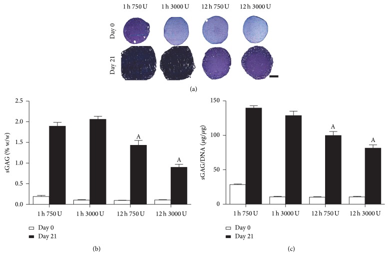
Figure 4.
sGAG accumulation of nasal chondrocytes isolated using digest protocols of 1 h or 12 h with 750 or 3000 U/ml of collagenase enzyme with physical agitation and subsequent culture in alginate beads for 21 days. (a) Histological evaluation with aldehyde fuchsin and alcian blue to identify sGAG at day 0 and day 21; deep blue/purple staining indicates sGAG accumulation and light blue staining indicates residual alginate. Scale bar: 1 mm (b). sGAG content normalized to percentage wet weight (% w/w) and (c) sGAG normalized on a per cell basis (sGAG/DNA). ASignificance to 1 h incubation for the same enzyme concentration, (p < 0.05).

