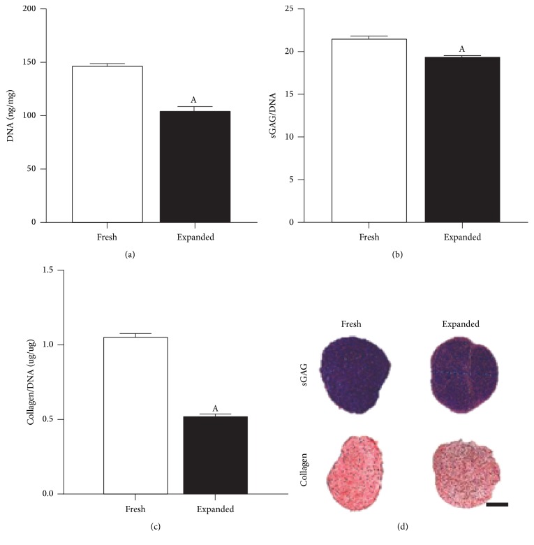
Figure 6.
Matrix forming capacity of freshly isolated (Fresh) and culture expanded (Expanded) chondrocyte pellet cultures after 21 days. Both populations were isolated using a 1 h rapid isolation protocol with 3000 U/ml of collagenase enzyme and physical agitation. “Fresh” chondrocytes were formed into pellets immediately after isolation with “Expanded” chondrocytes being subjected to 7 days of amplification on tissue culture plastic prior to pellet culture. (a) DNA content normalized to wet weight (ng/mg) at day 21. (b) sGAG normalized on a per cell basis (sGAG/DNA). (c) Collagen normalized on a per cell basis (Collagen/DNA). ASignificance compared to “Fresh” group, (p < 0.05). (d) Histological evaluation with aldehyde fuchsin and alcian blue to identify sGAG and picrosirius red to identify collagen deposits. Scale bar: 1 mm.

