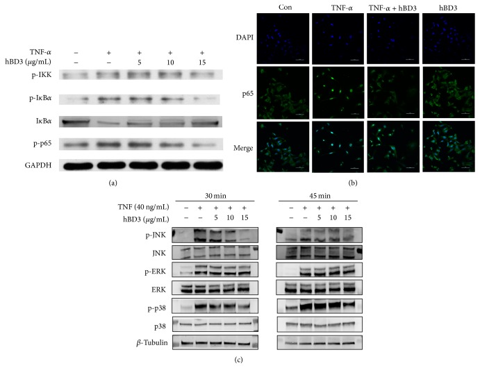
Figure 5.
Effects of hBD3 on TNF-α-induced NF-κB and MAPK activation in HUVECs. (a) HUVECs were incubated with TNF-α (40 ng/mL) with or without the indicated concentrations of hBD3 for 30 min. Whole cell lysates were centrifuged and analyzed with western blot using specific antibodies. (b) Immunofluorescence analysis for NF-κB p65 localization was conducted as mentioned above, and images were captured using a confocal laser scanning microscope system (Nikon A1, Japan) (original magnification: 400x, scale bar 50 μm). (c) HUVECs were incubated with TNF-α (40 ng/mL) with or without the indicated concentrations of hBD3 for 30 min and 45 min. Whole cell lysates were centrifuged and analyzed with western blot using specific antibodies.

