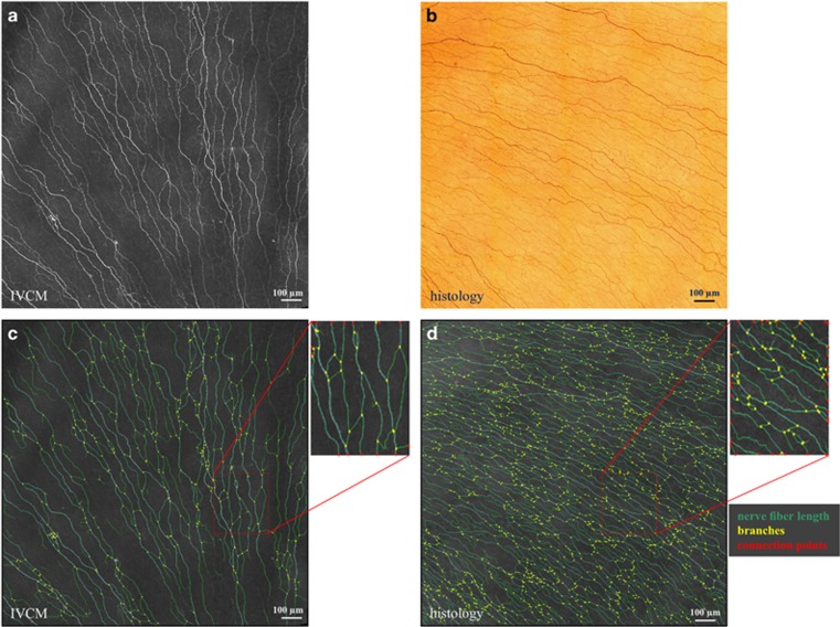
Figure 1.
SBP in images from IVCM and histology. (a) IVCM mosaic image of the human corneal SBP depicting corneal nerve fibers; (b) histological staining of the human corneal SBP showing β-III- tubulin-stained corneal nerves; and (c and d) represent the results of segmentation from the corresponding SBP images, respectively. Scale bar, 100 μm.

