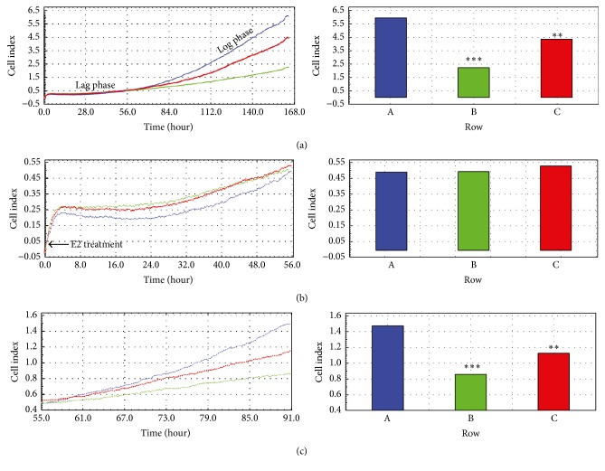
Figure 8.
Cell proliferation index (CI) of porcine follicular granulosa cells cultivated for 168 h after acute and prolonged E2 treatment. Porcine follicular granulosa cells were recovered from pubertal gilts and treated with collagenase for 10 min at 38.5°C. The cells were immediately transferred into an E-Plate 48 of a real-time cell analyzer (RTCA, Roche-Applied Science, GmbH, Penzberg, Germany). The experiment consisted of eight replicates involving the cultivation of the same population of collected cells. In the first experiment, the porcine GCs were cultured with 1.8 and 3.6 μM E2 at the beginning of an experiment (0 h), which was divided into three time periods: 0–168 h (Figure 8(a)), 0–56 h (Figure 8(b)), and 56–90 h (Figure 8(c)). The differences were considered to be significant at the level of ∗∗P < 0.01 and ∗∗∗P < 0.001. A, B, and C represent triplicates (three samples of the same population of collected cells).

