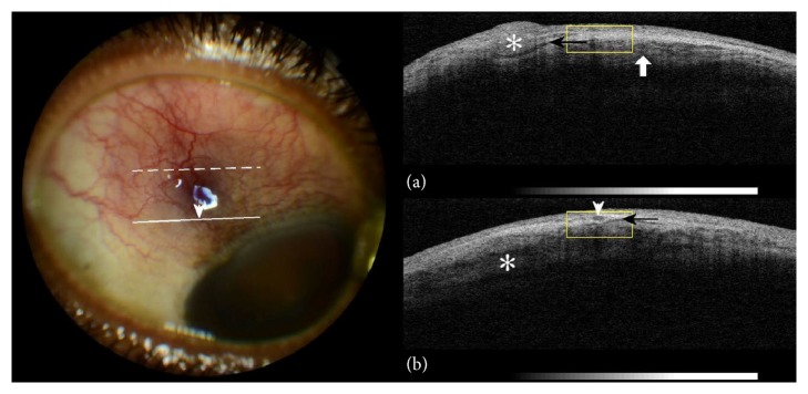
Figure 1.
On the left side: colour photography of the right eyeball; congestion of the superficial blood vessels and dilation of the episcleral vessels (arrowhead). On the right side: (a) anterior segment OCT (dashed line on eyeball photography). Nodular episcleral thickening (asterisk), subepiscleral fluid level (black arrow), and intralamellar scleral oedema (white arrow). (b) Anterior segment OCT (solid line on eyeball photography). Transverse section of dilated episcleral vessel (arrowhead) corresponding to relevant location on eyeball photography. Subepiscleral fluid level (black arrow) and intralamellar scleral oedema (asterisk).

