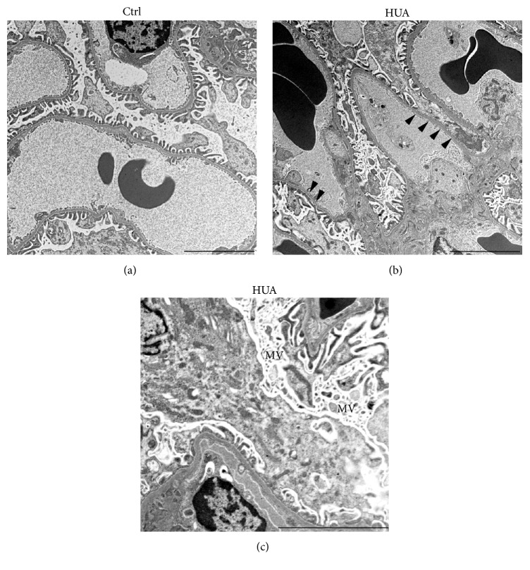
Figure 4.
Podocyte injury in hyperuricemic rats is confirmed by electron microscopy. Transmission electron micrographs of podocyte foot process in the glomeruli of indicated animals. Podocytes in the kidney from hyperuricemic rats (HUA) showed foot process effacement (arrowheads) and microvillus transformation (MV). These changes were less evident in the control (Ctrl) group. Bars represent 5 μm.

