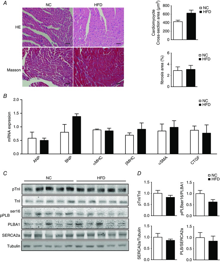
Figure 2. Mice after 8 weeks of HFD feeding display normal cardiac gene expression and morphology.

A, heart sections from NC and HFD mice were stained with Haematoxylin and Eosin (HE; scale bar = 100 μm) to examine heart morphology and stained with Masson's trichrome (scale bar = 100 μm) to examine fibrosis. The cardiomyocyte cross‐sectional areas and fibrosis‐positive areas were quantified and plotted (n = 3). B, RT‐PCR showing left ventricular expression of genes involved in cardiac hypertrophy and fibrosis in mice after 8 weeks of HFD. C and D, Western blots showing protein kinase A (PKA) phosphorylation of phospholamban (PLB) and troponin I (TnI), and SERCA2a expression in NC and HFD mouse hearts. [Color figure can be viewed at wileyonlinelibrary.com]
