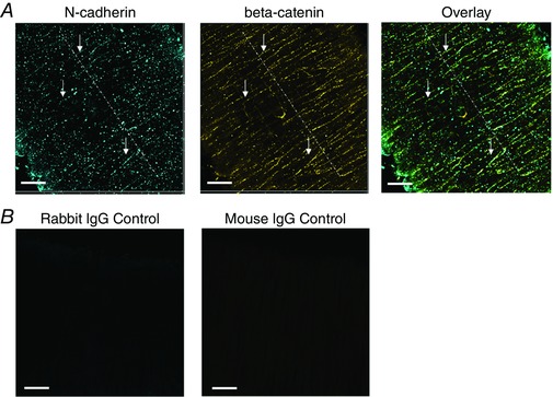
Figure 1. N‐cadherin forms AJs between VSMC.

A, N‐cadherin clusters are observed along the VSMC borders between adjacent cells in the vessel wall of SCAs. Many N‐cadherin and β‐catenin clusters appear patterned as a string of beads along cell–cell borders (depicted by white arrows). The SCA was pressurized to 90 mmHg and had myogenic tone. The vessel was fixed under pressure, immunolabelled for N‐cadherin and β‐catenin, and visualized by confocal microscopy. B, vessel labelled with control antibodies. The SCA was pressurized to 90 mmHg and had myogenic tone. The vessel was fixed under pressure, and immunolabelled with rabbit control IgG and mouse control IgG, followed by labelling with the same secondary antibodies. Scale bar = 20 μm. [Color figure can be viewed at wileyonlinelibrary.com]
