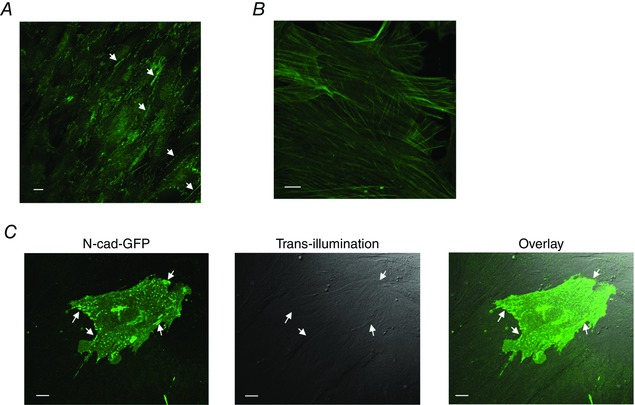
Figure 4. Both endogenous N‐cadherin and N‐cadherin‐EGFP were recruited to AJs in cultured VSMC.

A, immunofluorescence labelling of cell endogenous N‐cadherin in primary cultured VSMCs isolated from rat SCAs. Through‐focus stack images were collected using confocal fluorescence microscopy, and the stacked images were summed together to show the presence of N‐cadherin adherens junctions (white arrows). B, immunofluorescence labelling of smooth muscle α‐actin in primary cultured VSMCs. C, VSMCs were transfected with N‐cadherin‐EGFP. N‐cadherin‐EGFP was incorporated into adhesion structures between overlapping VSMCs. Left: a single cerebral VSMC in confluent culture exhibiting transfection with N‐cadherin‐EGFP was imaged using confocal fluorescence microscopy. White arrows point to the clusters of N‐cadherin‐EGFP. Middle: trans‐illumination image of the same area showing the transfected VSMC and neighbouring VSMCs that are not expressing N‐cadherin‐GFP. The neighbouring cells were overlapping and in contact with the cell expressing N‐cadherin‐EGFP. Right: overlay of the fluorescence image and trans‐illumination image. White arrows indicate the adhesion structures that incorporated N‐cadherin‐EGFP. Scale bar = 10 μm. [Color figure can be viewed at wileyonlinelibrary.com]
