FIG 1 .
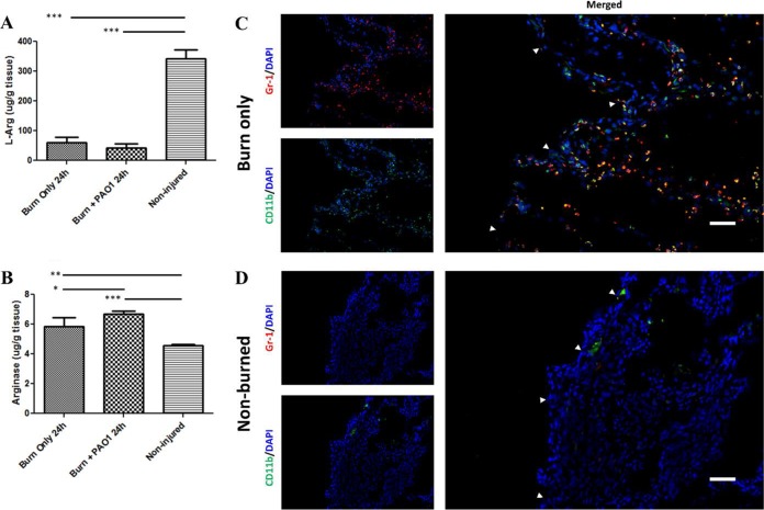
Depleted arginine levels are associated with MDSC recruitment. Tissue from mice that received thermal insult (with or without infection) was harvested 24 h post-burn injury. (A) l-Arg concentrations were significantly reduced in burned mice (with or without PAO1 infection) compared to the native concentrations of l-Arg in noninjured tissue by one-way analysis of variance (ANOVA) with Newman-Keuls multiple comparison posttest. ***, P < 0.001 (n = 5 mice/group). (B) Arginase concentrations were significantly elevated in tissue from mice that received a thermal insult or insult coupled with PAO1 infection compared to basal arginase levels in noninjured tissue by one-way ANOVA with Newman-Keuls multiple comparison posttest. ***, P < 0.001; **, P < 0.01; *, P < 0.05 (n = 5 mice/group). Tissue was harvested from (C) burn-only mice 24 h post-thermal insult or (D) nonburned mice. Tissue samples were prepared for direct immunofluorescence microscopy with FITC-labeled anti-CD11b and PE-labeled anti-Gr-1 antibodies. Host cell nuclei were counterstained with DAPI (blue). (C) A large number of MDSCs coexpressing both CD11b and Gr-1 are recruited to the burned tissue-intact tissue interface (white arrows). (D) No MDSCs were observed in the tissue from the dorsum of nonburned mice. Sections were visualized via a Nikon Plan Fluor 20×/0.75 objective. White size bars represent 50 µm.

